Book Appointment Now


Mammography Equipment: A Pivotal Innovation in the Timely Diagnosis of Breast Cancer
Breast cancer remains one of the most common cancers affecting women worldwide. Early detection is vital for successful treatment outcomes, and mammography equipment plays a pivotal role in this process. By providing detailed images of breast tissue, mammography allows healthcare professionals to identify abnormalities at their earliest stages. In recent years, advancements in mammography technology have enhanced its accuracy and accessibility, solidifying its importance in oncology.
How Mammography Equipment Works: A Detailed Description
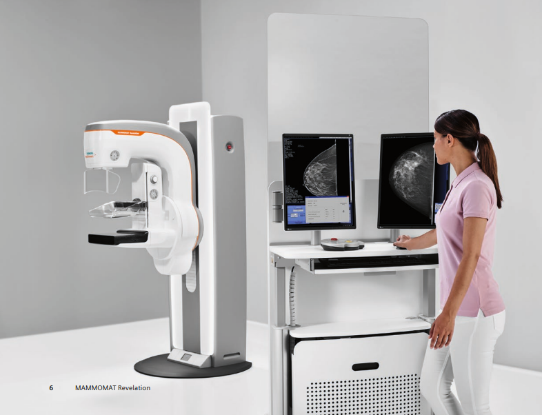
Mammography equipment is specialized imaging technology that uses low-dose X-rays to capture detailed images of breast tissue, known as mammograms. The main components of a mammography system include:
- X-ray Tube: Generates the X-rays needed to penetrate breast tissue.
- Compression Device: Gently compresses the breast to spread out the tissue for clearer images.
- Digital Detector or Film Cassette: Captures the X-ray images.
- Workstation: Processes and displays the images for analysis.
Mammography works by using X-rays, which are absorbed differently by various types of tissue. Dense tissues, like tumors, absorb more X-rays, appearing lighter on the image, while less dense tissues, like fat, absorb fewer X-rays and appear darker.
Modern mammography has largely transitioned from film to digital systems. With digital mammography, clinicians capture electronic images that allow them to manipulate, store, and share data more efficiently. This technology improves the detection of abnormalities, especially in women with dense breast tissue. These advanced systems not only improve image clarity but also reduce false positives in clinical evaluations.
Clinical Applications of Mammography Equipment in Oncology
Mammography is an indispensable tool in oncology, with a wide range of applications that extend beyond mere detection. Its role encompasses screening, diagnosis, treatment planning, and monitoring, making it a cornerstone in the comprehensive management of breast cancer.
1. Screening for Early Detection
Early detection of breast cancer significantly improves treatment outcomes and survival rates. Consequently, mammography has become the most effective and widely used screening method for this purpose.
- Routine Screening Programs: Health organizations recommend regular mammograms for women within certain age groups. For example, the American Cancer Society advises that women aged 40 to 54 should have annual mammograms, while those 55 and older can switch to biennial screening or continue yearly exams.
- Personalized Screening Schedules: Women with higher risk factors, such as a family history of breast cancer or genetic mutations (e.g., BRCA1 or BRCA2), may start screening earlier and undergo more frequent exams.
- Population Health Impact: Widespread screening has contributed to a decline in breast cancer mortality rates by enabling earlier detection and treatment.
2. Diagnostic Mammography
When abnormalities are detected during routine screening or when symptoms are present, diagnostic mammography is employed for a more detailed evaluation.
- Targeted Imaging: Provides additional views and magnifications to closely examine specific areas of concern, such as lumps, asymmetries, or unusual tissue densities.
- Characterizing Lesions: Helps distinguish between benign and malignant findings by analyzing shapes, margins, and patterns of calcifications.
- Guiding Clinical Decisions: Informs the need for further testing, such as biopsies or additional imaging modalities like ultrasound and MRI.
3. Using Mammography Equipment for Breast Cancer Treatment Planning
Moreover, accurate imaging is critical for developing effective treatment strategies tailored to the individual patient. Precision imaging is especially vital when assessing multifocal disease or planning targeted interventions.
- Assessing Tumor Size and Location: Precise measurements guide surgeons in deciding between breast-conserving surgery (lumpectomy) and mastectomy.
- Evaluating Multifocal and Multi centric Disease: Determines the presence of multiple tumors within the same breast, which may influence the extent of surgical intervention.
- Preoperative Localization: Radiologists use wire localization under mammographic guidance to mark non-palpable lesions and assist surgeons during excision.
4. How Mammography Equipment Helps Monitor Treatment Response
For patients undergoing neoadjuvant therapies (treatments given before surgery), mammography monitors how tumors respond to interventions. Thus, it helps in assessing tumor reduction and adjusting treatment plans.
- Assessing Tumor Reduction: Regular imaging evaluates changes in tumor size, indicating the effectiveness of chemotherapy or hormone therapy.
- Adjusting Treatment Plans: Results may lead oncologists to modify therapeutic approaches for better outcomes.
5. Post-Treatment Surveillance
After initial treatment, ongoing surveillance is essential to detect recurrences or new primary cancers. Therefore, routine follow-up mammograms are recommended.
- Routine Follow-Up Mammograms: Annual or biennial exams monitor the treated breast and the contralateral breast for any changes.
- Detecting Recurrence Early: Early identification of recurrent cancer can lead to prompt intervention and improved prognosis.
6. Guiding Interventional Procedures
Mammography equipment plays a foundational role not only in screening but also in guiding biopsies and surgical planning. In addition, mammography aids in minimally invasive diagnostic and therapeutic procedures.
- Stereotactic Breast Biopsy: Uses mammographic imaging to precisely locate and sample suspicious areas without the need for open surgery.
- Needle Localization: Assists in placing markers to guide surgeons during the removal of non-palpable abnormalities.
7. Special Applications
Furthermore, mammography is particularly adept at detecting tiny calcium deposits that may indicate early-stage cancers like ductal carcinoma in situ (DCIS).
- Assessing Micro calcifications: Identifies minute calcium deposits that may not be detectable through physical examination.
- Evaluating Breast Implants: Although MRI is the gold standard, mammography can sometimes identify implant ruptures or leaks.
- Male Breast Cancer Detection: While rare, breast cancer in men can be detected and evaluated using mammography.
8. Integration with Other Diagnostic Tools
Moreover, combining mammography with other imaging modalities enhances diagnostic accuracy.
- Ultrasound: Complements mammography, especially useful in evaluating dense breast tissue or distinguishing cysts from solid masses.
- Magnetic Resonance Imaging (MRI): Provides detailed images and is often used for high-risk patients or for further evaluation of ambiguous findings.
- Contrast-Enhanced Mammography: An emerging technique that improves visualization of tumor vascularity, aiding in the detection of aggressive cancers.
Example Scenario
A 48-year-old woman undergoes her annual screening mammogram. Modern mammography equipment enables clinicians to detect abnormalities that are often missed during physical exams. The radiologist notices a small area of architectural distortion in the upper outer quadrant of her right breast—a subtle abnormality not felt during a physical exam. A diagnostic mammogram with magnification views confirms the suspicion. An ultrasound-guided core needle biopsy is performed, and pathology reveals early-stage invasive ductal carcinoma. Because the cancer was detected at an early stage, the patient opts for a lumpectomy followed by radiation therapy. As a result, her prognosis is excellent, highlighting the critical role of mammography in early detection.
Impact on Different Patient Populations
Advancements in mammography equipment have led to better detection rates, particularly in women with dense breast tissue.
- Women with Dense Breasts: Dense breast tissue can obscure lesions, reducing mammography’s sensitivity. Therefore, supplemental screening methods like 3D mammography (tomosynthesis), ultrasound, or MRI may be recommended.
- High-Risk Individuals: Patients with genetic predispositions or prior chest radiation therapy may benefit from enhanced screening protocols, combining mammography with other imaging techniques.
9. Research and Clinical Trials
Additionally, mammography contributes to ongoing research aimed at improving breast cancer detection and treatment.
- Developing New Technologies: Clinical trials assess the efficacy of advanced imaging methods, such as tomosynthesis and AI algorithms.
- Epidemiological Studies: Large-scale data from mammography screenings help researchers understand cancer patterns and risk factors.
10. Artificial Intelligence and Mammography
Mammography equipment continues to evolve, integrating AI and digital technologies to improve diagnostic confidence. The integration of artificial intelligence marks a transformative step in mammography analysis and decision-making.
Moreover, AI is increasingly integrated into mammography to enhance diagnostic capabilities.
- Computer-Aided Detection (CAD): Algorithms analyze images to highlight areas that may require further review by a radiologist.
- Improving Accuracy: AI can reduce false positives and negatives by identifying subtle patterns not easily detected by the human eye.
- Workflow Efficiency: Automating initial image analysis can streamline radiology workflows, allowing clinicians to focus on more complex cases.
Public Health Considerations
Furthermore, public health initiatives enhance the overall impact of mammography. Portable mammography equipment is increasingly used in mobile screening units to reach underserved communities.
- Accessibility and Equity: Mobile mammography units and community outreach programs aim to improve access in rural and underserved areas.
- Education and Awareness: Public health campaigns promote awareness about the importance of regular screenings and breast health.
- Policy and Guidelines: Health organizations continuously evaluate screening recommendations to balance benefits with potential risks like over diagnosis.
Challenges and Limitations
However, mammography is not without its challenges.
- False Positives: Approximately 10% of women are recalled for additional testing after a screening mammogram, which can cause anxiety and lead to unnecessary procedures.
- False Negatives: Mammography misses about 20% of breast cancers, emphasizing the need for complementary screening methods in certain populations.
- Radiation Exposure: While doses are low, minimizing radiation exposure remains a priority, especially for women requiring frequent imaging.
Future Directions
Looking ahead, several initiatives aim to enhance the effectiveness of mammography.
- Personalized Screening: Tailoring screening protocols based on individual risk factors, including genetic profiles and breast density, is being considered.
- Technological Advancements: Development of low-dose mammography and enhanced imaging techniques like spectral mammography.
- Global Health Initiatives: Expanding access to mammography in developing countries through cost-effective technologies and training programs.
Advantages and Limitations
Advantages:
- Early Detection: Identifies cancers before symptoms develop, improving treatment success rates.
- Improved Survival Rates: Early-stage detection is linked to higher survival rates.
- Non-Invasive: Mammography is a non-invasive procedure with minimal discomfort.
Limitations:
- False Positives: May lead to unnecessary additional testing and anxiety.
- False Negatives: A small percentage of cancers may be missed, especially in dense breasts.
- Radiation Exposure: Although the radiation dose is low, there is nevertheless a minimal risk associated with repeated exposure.
Recent Developments and Innovations

Recent advancements have significantly enhanced mammography technology. This breakthrough in breast imaging has significantly improved the accuracy of early-stage tumor detection.
Digital breast tomosynthesis is revolutionizing how clinicians detect and assess abnormalities in dense breast tissue.
- Digital Breast Tomosynthesis (3D Mammography): Produces three-dimensional images by taking multiple X-ray pictures of the breast from different angles. Consequently, this method improves cancer detection rates and reduces false positives.
- Contrast-Enhanced Mammography: Involves injecting a contrast agent to highlight areas of increased blood flow, often associated with tumors.
- Artificial Intelligence Integration: AI algorithms assist radiologists by detecting patterns and anomalies, thereby improving diagnostic accuracy and efficiency.
- Portable Mammography Units: Increase accessibility in remote or underserved areas, facilitating broader screening efforts.
Ongoing research focuses on refining AI applications and enhancing image quality while reducing radiation doses.
Safety and Patient Comfort
Safety Features:
- Low Radiation Dose: Adheres to the “As Low As Reasonably Achievable” (ALARA) principle to minimize exposure.
- Regular Calibration and Maintenance: Ensures equipment operates safely and effectively. (Regular maintenance and calibration of mammography equipment are essential to ensure consistent image quality and patient safety.)
- Compliance with Standards: Meets regulatory guidelines set by organizations like the FDA and the American College of Radiology.
Patient Comfort Considerations:
- Ergonomic Design: Modern machines are designed to reduce discomfort during breast compression.
- Technologist Training: Professionals are trained to position patients properly and explain procedures to alleviate anxiety.
- Privacy and Support: Facilities provide a supportive environment to address patient concerns.
Patients are encouraged to communicate any discomfort or concerns to the technologist to improve the experience.
Conclusion
Mammography is not just an indispensable tool in oncology, it is a life-saving technology that plays a critical role in the early detection and diagnosis of breast cancer, offering patients the best chance for successful treatment.
Mammography continues to play a pivotal role in reducing mortality rates through early intervention and diagnosis. Looking forward, the ongoing evolution of mammography promises even greater improvements in cancer care, with potential developments in personalized screening protocols and further reductions in radiation exposure. Furthermore, global initiatives aim to make mammography more accessible, ensuring that more women benefit from early detection. As we advance, mammography will remain at the forefront of the fight against breast cancer, saving lives through early detection and intervention.
References:
- American Cancer Society. (2023). Breast Cancer Early Detection and Diagnosis.
- Radiological Society of North America. (2023). Mammography.
- World Health Organization. (2023). Breast Cancer: Prevention and Control.
For more information on cancer care and advancements in oncology equipment, visit CancersHub.com.


