Book Appointment Now


Digital X-Ray Machines in Oncology: Illuminating Cancer Diagnosis and Treatment
Introduction
Early and accurate detection is crucial in the fight against cancer. Digital X-ray machines have become indispensable tools in oncology, providing quick and detailed images that aid in diagnosing, staging, and monitoring various cancers. With advancements in digital technology, these machines offer enhanced image quality, reduced radiation exposure, and faster results compared to traditional film-based X-rays. Their increasing relevance in recent years has significantly improved patient care and outcomes in cancer treatment.
Detailed Description
Digital X-ray machines use electromagnetic radiation to capture images of the inside of the body. Unlike traditional film X-rays, digital X-rays convert the transmitted X-ray beams into digital signals, producing images that can be viewed immediately on a computer screen.
Key Components:
- X-ray Tube: Generates X-rays by accelerating electrons and colliding them with a metal anode.
- Digital Detector: Replaces traditional film; captures X-rays that pass through the body and converts them into digital images.
- Computer System: Processes and displays the images for analysis.
- Control Console: Allows the technician to adjust settings like exposure time and radiation dose.
- Patient Table or Stand: Positions the patient appropriately for the examination.
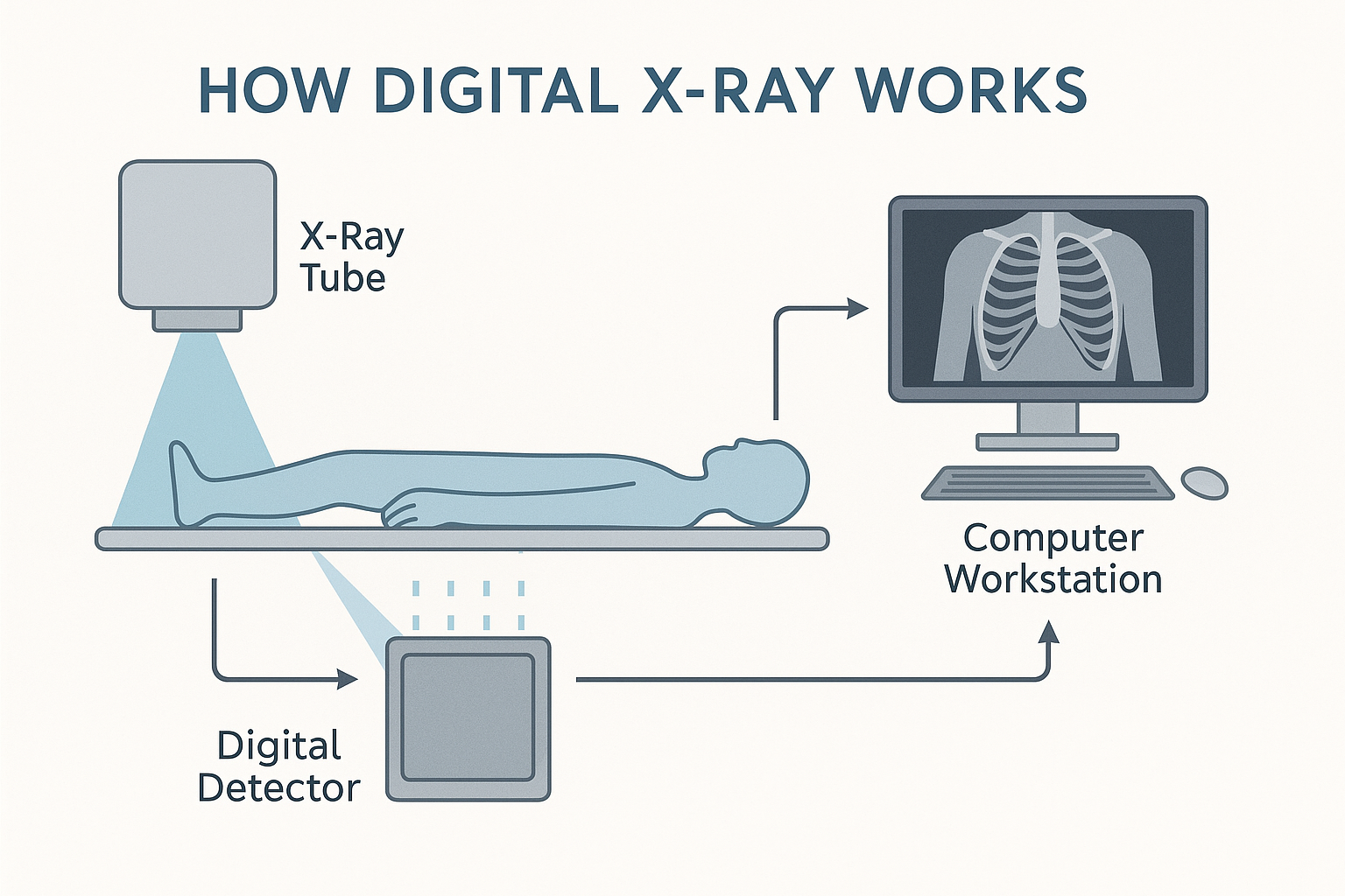
How It Works:
- Generation of X-rays: The X-ray tube produces X-rays when high-speed electrons collide with a metal target, emitting radiation.
- Transmission Through the Body: X-rays pass through the patient’s body and are absorbed differently by various tissues. Dense tissues like bone absorb more X-rays, appearing white on the image, while softer tissues absorb less and appear in shades of gray.
- Detection and Conversion: The digital detector captures the X-rays that pass through the body and converts them into electronic signals.
- Image Processing: The computer system processes these signals to create a digital image that can be enhanced, zoomed, and adjusted for better visualization.
Think of it like taking a digital photograph of the inside of the body, where bones and tumors can be seen clearly due to their density differences.
Applications in Oncology
Digital X-ray machines are versatile tools in cancer care, serving multiple roles:
1. Detection and Diagnosis
- Identifying Tumors: X-rays can reveal masses or abnormalities in organs such as the lungs, bones, and breasts.
- Screening: Used in mammography for early detection of breast cancer.
- Evaluating Symptoms: Helps investigate unexplained pain, fractures, or persistent cough.
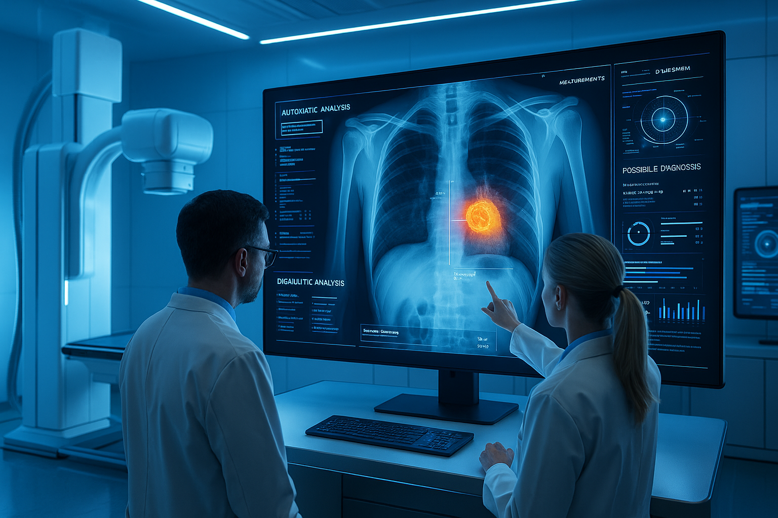
Scenario:
A patient complains of persistent chest pain and coughing. A digital chest X-ray reveals a suspicious mass in the lung. Early detection leads to further testing, confirming a diagnosis of lung cancer, and allowing prompt initiation of treatment.
2. Staging and Monitoring
- Assessing Cancer Spread: Determines if cancer has spread to bones or other areas.
- Monitoring Treatment Effects: Regular X-rays track changes in tumor size or bone healing after therapy.
3. Guiding Procedures
- Biopsy Assistance: Helps in guiding needles for tissue sampling.
- Treatment Planning: Assists in preparing for surgeries or radiation therapy by providing detailed images of the affected area.
4. Post-Treatment Surveillance
- Detecting Recurrence: Monitors patients after treatment to ensure cancer has not returned.
- Evaluating Complications: Identifies issues like fractures or infections that may arise during cancer treatment.
Advantages and Limitations
Advantages:
- Immediate Results: Digital images are available instantly, reducing waiting times.
- Enhanced Image Quality: Ability to adjust contrast and brightness for better visualization.
- Reduced Radiation Exposure: Advanced detectors require lower doses of radiation compared to traditional X-rays.
- Easy Storage and Sharing: Digital images can be stored electronically and shared with other healthcare providers quickly.
- Cost-Effective: Generally less expensive than other imaging modalities like CT or MRI.
Limitations:
- Radiation Exposure: Although reduced, there is still exposure to ionizing radiation.
- Limited Soft Tissue Contrast: Less effective in imaging soft tissues compared to MRI or CT scans.
- Overlapping Structures: Two-dimensional images can sometimes make it difficult to interpret complex anatomical areas.
- Less Sensitivity for Early Detection: May not detect very small tumors or early-stage cancers without additional imaging techniques.
Recent Developments and Innovations
1. Digital Tomosynthesis
- Feature: Creates a 3D image of the body by taking multiple X-ray images from different angles.
- Benefit: Improves detection of small tumors, particularly in breast cancer screening (3D mammography).
2. Advanced Detectors
- Feature: Use of flat-panel detectors enhances image resolution.
- Benefit: Provides clearer images with lower radiation doses.
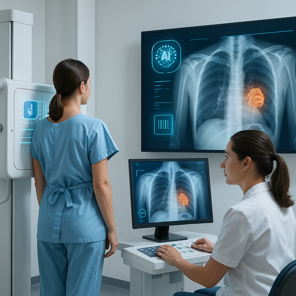
3. Artificial Intelligence Integration
- Feature: AI algorithms assist in image analysis.
- Benefit: Improves accuracy in detecting abnormalities and reduces diagnostic errors.
4. Portable Digital X-ray Machines
- Feature: Compact, mobile units that can be brought to the patient’s bedside.
- Benefit: Increases accessibility for patients who cannot be easily moved, such as those in intensive care.
5. Dose Reduction Technologies
- Feature: Advanced software optimizes image quality while minimizing radiation exposure.
- Benefit: Enhances patient safety, especially important for children and patients requiring multiple scans.
Ongoing Research:
- Dual-Energy X-ray Imaging: Captures images at two different energy levels to improve tissue characterization.
- Contrast-Enhanced Digital Mammography: Uses contrast agents to highlight areas of increased blood flow, aiding in cancer detection.
Safety and Patient Comfort
Safety Features:
- Radiation Dose Management: Machines are designed to use the lowest possible radiation dose to achieve quality images.
- Protective Measures: Use of lead aprons and shields to protect parts of the body not being imaged.
- Regular Maintenance and Calibration: Ensures equipment operates safely and effectively.
- Compliance with Standards: Adheres to regulations set by organizations like the FDA and the International Atomic Energy Agency (IAEA).
Patient Comfort Considerations:
- Quick Procedures: Typically completed within minutes, reducing discomfort.
- Non-Invasive: Painless with no need for injections (unless contrast agents are used).
- Comfortable Positioning: Technologists assist patients in finding a comfortable position for the best image quality.
- Clear Communication: Patients are informed about the procedure, helping to alleviate anxiety.
Addressing Patient Concerns:
- Radiation Exposure: Technologists explain that the benefits outweigh the minimal risks and all precautions are taken to minimize exposure.
- Pregnancy Considerations: Special care is taken to avoid unnecessary exposure during pregnancy; alternative imaging methods may be recommended.
Conclusion
Digital X-ray machines have revolutionized cancer care by providing fast, accurate, and detailed images essential for diagnosis, treatment planning, and monitoring. Their ability to produce immediate results with enhanced image quality has significantly improved patient outcomes. Advancements like digital tomosynthesis and AI integration continue to enhance their capabilities, making them even more valuable in oncology.
Looking ahead, ongoing innovations promise to further improve image resolution, reduce radiation doses, and expand diagnostic applications. Digital X-ray technology will continue to be a cornerstone in cancer care, offering hope for earlier detection and more effective treatments, ultimately improving patient quality of life.
References
- American Cancer Society. (2023). Understanding X-rays for Cancer Diagnosis.
- Radiological Society of North America. (2023). Digital Radiography (Digital X-ray).
- National Cancer Institute. (2023). X-rays and Cancer Treatment.
For more information on cancer care and advancements in oncology equipment, visit CancersHub.com.
Feel free to learn more about digital X-ray machines and other medical equipment transforming cancer diagnosis and treatment. Being informed can empower patients and their families during their healthcare journey.


