Book Appointment Now

Breast Ultrasound Equipment: A Vital Tool in the Fight Against Breast Cancer
Introduction
Breast cancer is a leading cause of cancer-related deaths among women worldwide. Early detection and accurate diagnosis are crucial for improving survival rates. Breast ultrasound equipment plays an indispensable role in this process by providing real-time images of breast tissue. It complements other imaging modalities like mammography, especially in women with dense breast tissue.
In recent years, advancements in ultrasound technology have enhanced its effectiveness, making it an increasingly important tool in oncology.
Detailed Description
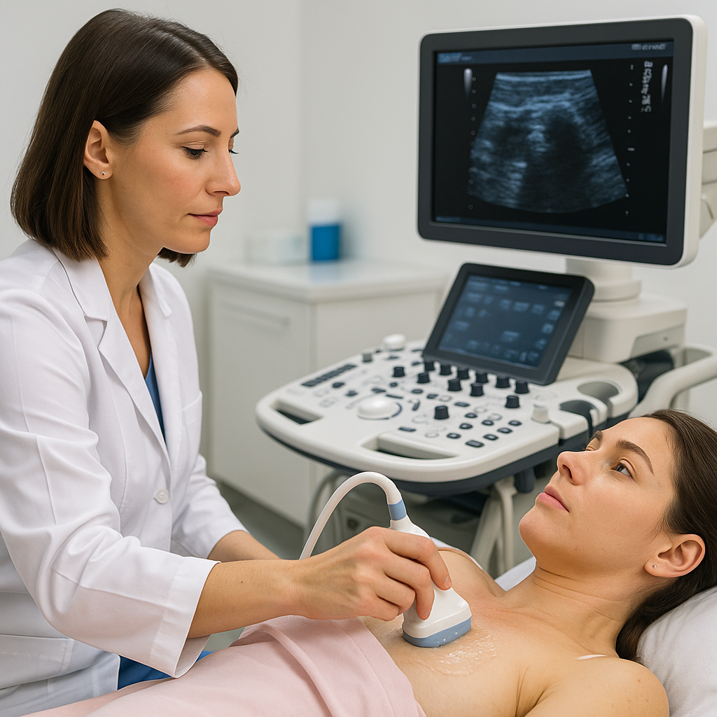
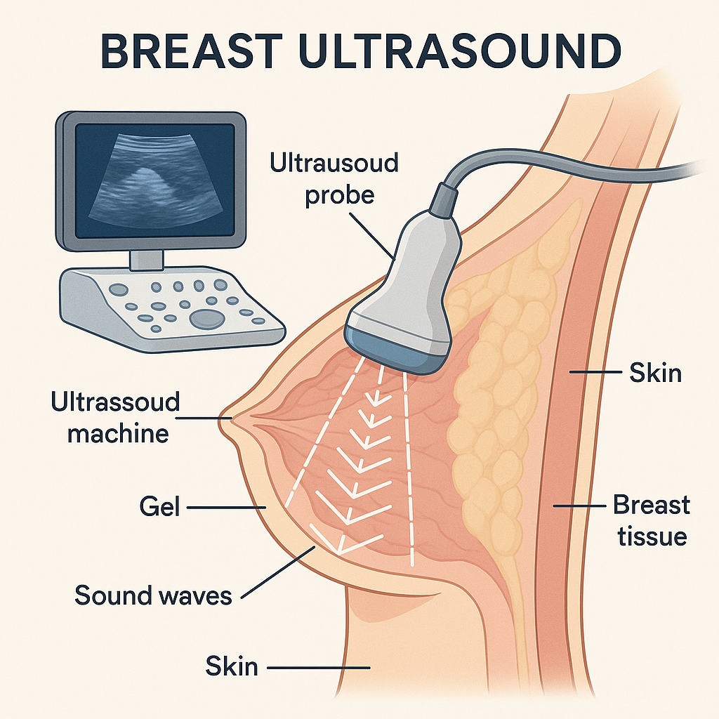
Breast ultrasound equipment utilizes high-frequency sound waves to produce images of the internal structures of the breast. The primary components of a breast ultrasound system include:
- Transducer (Probe): Emits sound waves and receives the echoes reflected back from tissues.
- Ultrasound Machine: Processes the echoes to create visual images displayed on a monitor.
- Gel: Applied to the skin to eliminate air pockets and facilitate the transmission of sound waves.
The fundamental principle behind ultrasound imaging is the reflection of sound waves. When the transducer is placed on the skin, it sends sound waves into the breast tissue. Different types of tissues reflect these waves back at varying speeds and intensities. The machine then interprets these echoes to construct a real-time image of the breast’s internal structures.
Because ultrasound does not use ionizing radiation, it is considered safe for repeated use. Modern breast ultrasound systems may also incorporate Doppler technology to assess blood flow within the breast tissue, which can be indicative of tumor activity.
Applications in Oncology
Breast ultrasound has a wide range of applications in oncology, extending from detection to treatment monitoring. Its versatility makes it an essential tool in comprehensive cancer care.
1. Diagnostic Evaluation
When abnormalities are detected through physical examination or other imaging modalities, ultrasound is often used for further evaluation.
- Characterizing Masses: Ultrasound helps distinguish between solid tumors and fluid-filled cysts.
- Assessing Lesion Margins: Determines whether a mass has well-defined or irregular edges, aiding in malignancy assessment.
- Guiding Biopsies: Real-time imaging allows for precise needle placement during minimally invasive biopsy procedures.
Scenario
A 40-year-old woman discovers a lump during a self-examination. A mammogram reveals dense breast tissue, making it difficult to assess the area thoroughly. An ultrasound is performed, which identifies a solid mass with irregular borders. Guided by the ultrasound, a biopsy is conducted, confirming the presence of malignant cells. Early detection enables prompt treatment, significantly improving her prognosis.
2. Screening in Dense Breast Tissue
In women with dense breast tissue, mammography’s sensitivity decreases. Ultrasound serves as an effective supplementary screening tool.
- Enhanced Detection: Improves the identification of small cancers that mammography may miss.
- Non-Ionizing Imaging: Offers a safe alternative without additional radiation exposure.
3. Monitoring Treatment Response
Ultrasound is valuable for tracking changes in tumor size during chemotherapy or other treatments.
- Real-Time Assessment: Provides immediate feedback on treatment efficacy.
- Adjusting Treatment Plans: Enables oncologists to modify therapies based on tumor response.
4. Guidance for Interventional Procedures
Ultrasound assists in various interventional oncology procedures.
- Needle Localization: Aids in placing markers before surgery.
- Ablation Therapy Guidance: Helps direct energy-based treatments that destroy tumor tissue.
5. Evaluating Axillary Lymph Nodes
Assessing lymph node involvement is critical in breast cancer staging.
- Detection of Metastasis: Identifies enlarged or suspicious lymph nodes under the arm.
- Biopsy Guidance: Facilitates fine-needle aspiration of lymph nodes for pathological examination.
6. Differentiating Benign from Malignant Lesions
Ultrasound elastography measures tissue stiffness, assisting in distinguishing benign from malignant masses.
- Elastography Applications: Stiffer tissues often indicate malignancy, aiding diagnostic accuracy.
- Complementary Data: Provides additional information alongside traditional imaging.
Advantages and Limitations
Advantages
- No Radiation Exposure: Safe for all patients, including pregnant women.
- Real-Time Imaging: Allows for immediate assessment and intervention.
- Cost-Effective: Generally less expensive than other imaging modalities.
- Portable Equipment: Can be used in various settings, including bedside examinations.
- Effective in Dense Breasts: Overcomes limitations of mammography in dense tissue.
Limitations
Not a Standalone Screening Tool: Generally used in conjunction with mammography rather than replacing it.
Operator Dependency: Image quality and interpretation heavily rely on the technician’s skill.
Limited Field of View: May miss lesions located deep within the breast or near the chest wall.
False Positives: Can lead to unnecessary biopsies due to difficulty distinguishing some benign and malignant lesions.
Recent Developments and Innovations
Recent advancements have significantly enhanced the capabilities of breast ultrasound equipment.
- Automated Breast Ultrasound Systems (ABUS): Provide 3D images of the entire breast, reducing operator dependency and improving reproducibility.
- Ultrasound Elastography: Measures tissue stiffness, aiding in the differentiation of benign and malignant lesions.
- Contrast-Enhanced Ultrasound: Involves using microbubble contrast agents to highlight tumor vascularity.
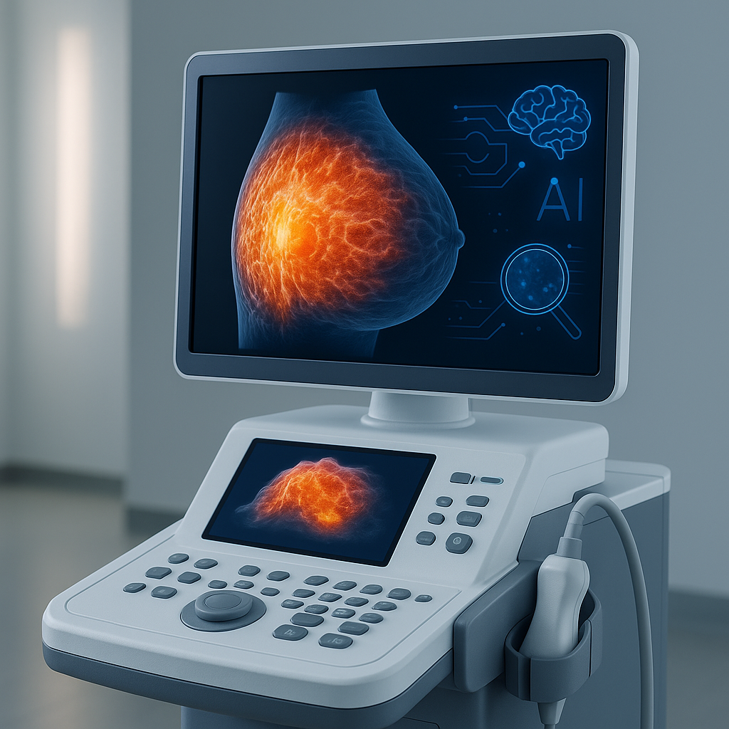
- Artificial Intelligence Integration: AI algorithms assist in image interpretation, improving diagnostic accuracy and reducing false positives.
- Portable and Handheld Devices: Increase accessibility, especially in remote or underserved areas.
Ongoing research focuses on refining AI applications and integrating ultrasound data with other imaging modalities for comprehensive analysis.
Safety and Patient Comfort
Safety Features
- Non-Ionizing Radiation: Uses sound waves, eliminating the risks associated with X-rays.
- Regulatory Compliance: Equipment meets standards set by organizations like the FDA and IEC.
- Regular Maintenance: Ensures devices operate safely and effectively.
Patient Comfort Considerations
- Non-Invasive Procedure: Generally painless and does not require injections or incisions.
- Comfortable Environment: Examinations are typically conducted in a private, relaxed setting.
- Flexible Positioning: Patients can often be scanned in comfortable positions.
Patients are encouraged to communicate any discomfort or concerns during the procedure to the technologist.
Conclusion
Breast ultrasound equipment is a vital tool in oncology, offering a safe, effective means of detecting and evaluating breast abnormalities. Its ability to provide real-time, detailed images without radiation exposure makes it an invaluable complement to mammography, particularly for women with dense breast tissue. Advancements in technology, such as automated systems and AI integration, continue to enhance its diagnostic capabilities.
Looking forward, the role of breast ultrasound in cancer care is expected to expand further. Innovations aimed at improving accuracy and accessibility will likely make it an even more integral part of early detection and personalized treatment strategies. By staying at the forefront of technological advancements, breast ultrasound will continue to play a critical role in improving patient outcomes and saving lives.


